Die Vielfalt GABAerger inhibitorischer Synapsen im gesunden und kranken Gehirn
GABAerge Schaltkreise regulieren den Informationsfluss im Gehirn und beeinflussen dadurch Verhalten. Störungen der GABAergen Signalübertragung treten bei zahlreichen psychiatrischen und neurologischen Erkrankungen auf. Zwar werden GABA(A)-Rezeptoren bereits pharmakologisch behandelt, doch wirken viele Medikamente unspezifisch im gesamten zentralen Nervensystem und verursachen dadurch relevante Nebenwirkungen.

Unsere Arbeitsgruppe untersucht daher die molekulare Vielfalt GABAerger Synapsen. Diese unterscheiden sich je nach Hirnregion und Neuronentyp und könnten dadurch gezieltere therapeutische Ansätze ermöglichen. Durch molekulare, funktionelle und verhaltensbezogene Untersuchungen in Mausmodellen wollen wir schaltkreisspezifische Zielstrukturen identifizieren. Im Fokus stehen GABAerge Synapsen in Amygdala, Hippocampus und präfrontalem Kortex sowie ihre Bedeutung für Angst- und Sozialverhalten.


aktuelle Projekte

Functional characterization of Nlgn2 at different GABAergic synapse subtypes in the mouse brain

Tamara Ritter, M.Sc.

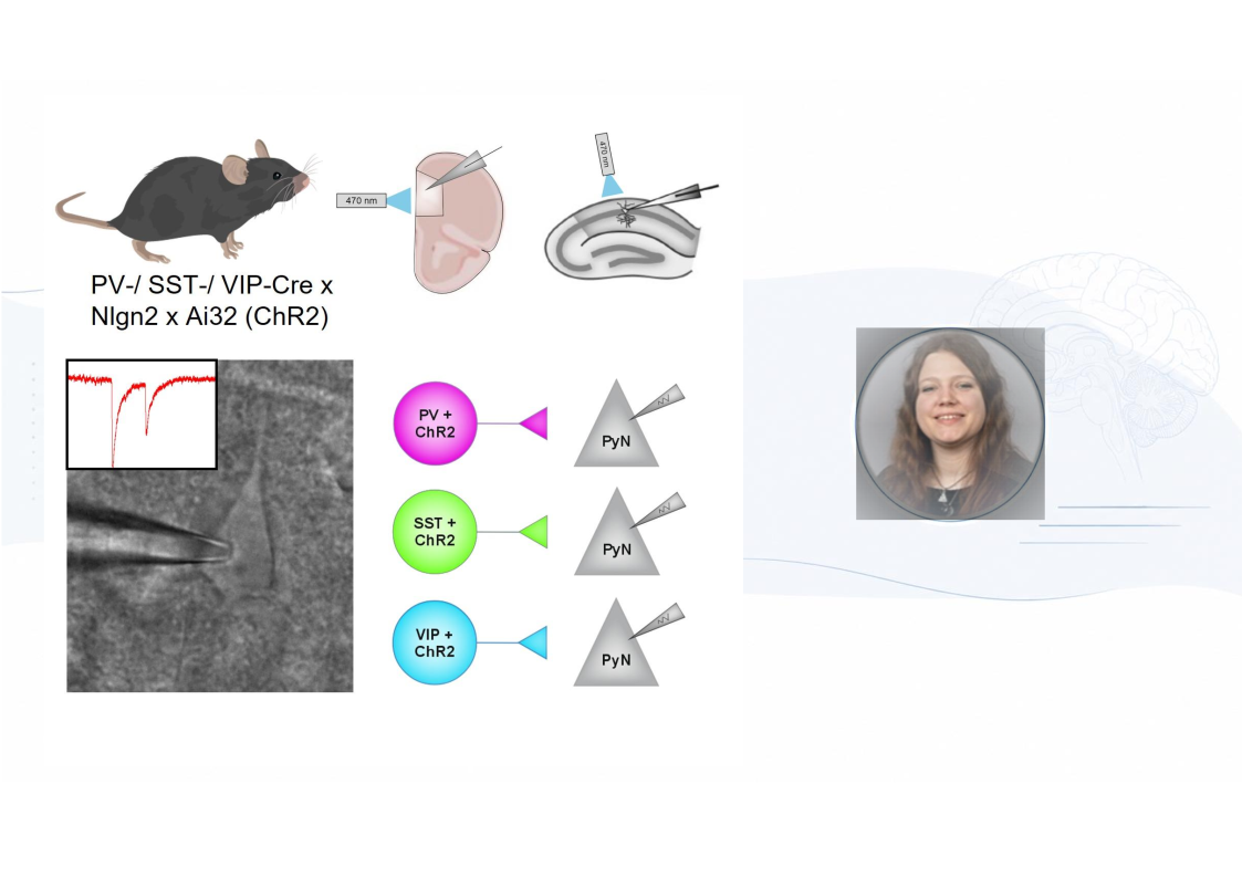
Dysfunction of inhibitory transmission disturbs the balance of excitation and inhibition (E/I), which contributes to neurological and psychiatric disorders. Despite its importance, the underlying mechanisms leading to E/I imbalance are poorly understood. GABA receptors of type A (GABAARs) are key regulators of neuronal activity and are recruited and anchored at GABAergic synapses by organizer complexes. The latter contain transsynaptic adhesion proteins such as Neuroligin2 (Nlgn2). Nlgn2 mutations have been shown to impair inhibitory synaptic transmission and lead to GABAAR clustering as well as psychiatric disorders. However, the specific distribution and precise function of Nlgn2 at different GABAergic synapse subtypes of various brain regions are yet to be understood. In this context two brain regions are of major interest: the medial prefrontal cortex (mPFC) and the hippocampal CA1 region. Impairments of these regions have been associated with a variety of neurological and psychiatric disorders. Moreover, recent findings indicated a distinct function of Nlgn2 at different GABAergic synapse subtypes in the CA1 region compared to cortical regions. To fill this gap, this study aimed to compare the function of Nlgn2 at parvalbumin (PV), somatostatin (SST) and vasoactive intestinal peptide (VIP) expressing GABAergic synapse subtypes in the mPFC and CA1 region. This is analyzed by applying focal light stimulation to specific presynaptic neuron types expressing ChR2 and simultaneously recording postsynaptic responses of connected neurons. To understand how Nlgn2 shapes neuronal circuits in health and disease, it is critical to resolve the longstanding question at which GABAergic synapses Nlgn2 exerts its effect.

GABAergic synapse subtype-specific effects of existing and future GABAergic pharmacotherapies in mice

Kyra Sohns, PhD

The synapse is the port of information transfer between nerve cells in the brain. The balance between excitatory and inhibitory synapses is crucial for regulating excitability within the nervous system and is essential to correct information processing within the brain. In recent years, inhibitory synapses in particular have become an important research focus. Elucidating the molecular mechanisms regulating the properties of GABAergic synaptic neurotransmission is of fundamental importance for understanding brain function in health and disease.

One important feature to be considered when thinking of GABAergic synapses as therapeutic targets is their variety. They are defined both by the identity of the presynaptic inhibitory neuron and by the components of the postsynaptic complex (Krueger-Burg, 2025). It is well-known that the various subtypes of presynaptic inhibitory neurons play very different roles in neural circuits and thus influence psychiatrically-relevant behaviours in different ways. In contrast, the role played by the various postsynaptic GABAAR subtypes and organizational proteins in circuits relevant to neuropsychiatric disorders, and how they relate to the diversity of presynaptic inhibitory neurons, remains largely unknown. Investigation into the architecture of individual inhibitory synapses and the overarching organisational principals of inhibitory connectivity in the brain could allow us specifically target GABAAR subtypes in circuits relevant to psychiatric disorders pharmacologically and thus avoid serious potential side effects that some currently-used pharmacotherapies present. 

The aim of this study is to investigate whether existing GABAergic pharmacotherapies exhibit specificity for certain synaptic subtypes in the medial prefrontal cortex (mPFC) and hippocampus (HPC) of mice. Using electrophysiology alongside a viral construct and immunohistochemistry, we aim to characterize the synaptic effects of pharmacotherapies and identify which components are present at specific GABAergic synaptic subtypes, thereby clarifying if certain GABAergic drugs more effectively target particular subtypes, which could pave the way for more targeted therapies with fewer side effects.
Early-life stress, microglia, and inhibitory circuits

Juliette Soubra, M.Sc.

Early postnatal development is a critical period of plasticity during which experience shapes neural circuits. Chronic stress, neglect, or abuse during this period (ie, early life stress) can produce lasting changes that increase the risk of anxiety and other psychiatric disorders. Understanding this vulnerability requires identifying which developing circuits are altered.

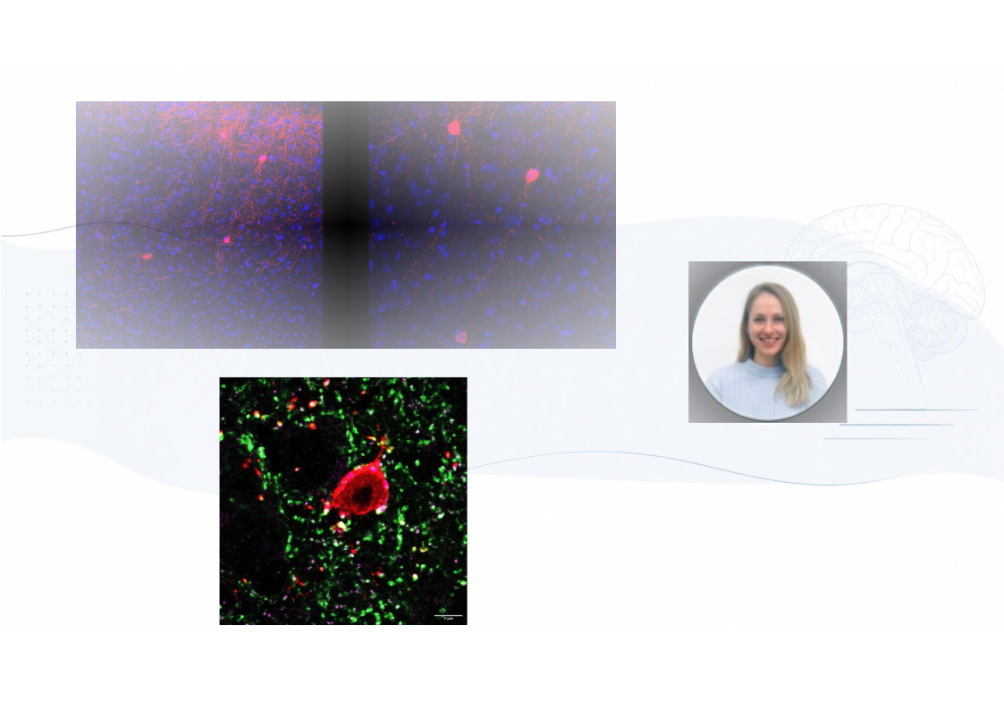
Anxiety involves the corticolimbic circuit comprising the amygdala, the ventral hippocampus, and the medial prefrontal cortex. Its function depends on a balance between excitation and inhibition, maintained in part by GABAergic interneurons. Parvalbumin interneurons regulate neuronal output and network synchrony, whereas somatostatin interneurons control the integration of excitatory inputs along dendrites. Early-life stress may disrupt these populations differently, biasing the network toward excessive threat responses. Microglia may mediate this disruption. In addition to their role as the immune cells of the brain, microglia prune synapses during development in response to neuronal activity and stress. Altered microglial interactions with inhibitory synapses during a critical developmental period could therefore weaken GABAergic control and produce persistent changes in the corticolimbic circuit.

The central hypothesis is that microglia could regulate developing inhibitory synapses in an interneuron-specific manner and that early-life stress disrupts this regulation. The project tests this hypothesis through three objectives:

  1. Determine how microglia interact with inhibitory synapses. Immunohistochemistry, electrophysiology, and optogenetics in mice will assess microglial interactions with synapses formed by specific interneuron populations and their effects on inhibitory transmission across corticolimbic regions.
  2. Establish how early-life stress alters this regulation. A mouse model of early-life stress will be used to investigate whether changes in microglial synaptic remodelling are accompanied by impaired GABAergic circuit function.
  3. Identify how interneuron activity communicates with microglia.Chemogenetic manipulation, imaging, and molecular profiling will determine how the activity of specific interneuron populations drives microglial responses.

Together, these experiments could explain how early life stress impacts developing circuits and leads to the development of anxiety disorders.

The role of VIP-expressing neurons in neuropsychiatrically relevant behaviours

Marina Taylor, M.Sc.

The GABAergic inhibitory system regulates neuronal excitability, network activity, and excitation–inhibition balance, yet its dysfunction in neuropsychiatric disorders remains poorly understood (Marín, 2012). This project focuses on vasoactive intestinal peptide-expressing (VIP⁺) interneurons, which modulate cortical and limbic circuits through disinhibition. Although emerging evidence links VIP+ neuron dysfunction to neurodevelopmental and psychiatric disorders, its consequences for anxiety-like, social, and fear-related behaviours remains poorly understood (Mossner et al., 2020; Zhu et al., 2025).

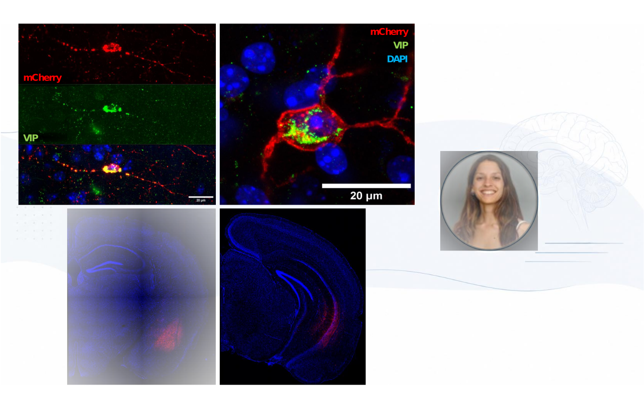
Therefore, this project aims to determine the contribution of VIP-expressing neurons to behaviours relevant to psychiatric disorders. To this end, VIP⁺ neurons were chemogenetically activated or inhibited in the medial prefrontal cortex (mPFC), basolateral amygdala (BLA), and ventral hippocampus (vHPC), three corticolimbic regions implicated in psychiatric disorders. Following VIP⁺ neuron manipulation, social behaviour, anxiety-related behaviour, associative fear learning, and fear expression were assessed. Chemogenetics was selected because it induces sustained changes in neuronal excitability lasting several hours, providing a model that more closely reflects the persistent circuit dysfunction associated with psychiatric disorders than brief, transient manipulations.

Our findings reveal a strikingly region- and behavior-specific profile: only sustained manipulation of mPFC VIP⁺neurons impaired behavior and exclusively in fear-related domains. No effects were observed in social or anxiety-related behaviors, nor were comparable deficits seen following VIP⁺ modulation in the BLA or vHPC. These results position mPFC VIP⁺ neurons as selective regulators of fear circuits. Their functional specificity makes them compelling translational targets. Future work must identify the molecular effectors governing mPFC VIP⁺ function, with the long-term goal of developing precision therapeutics that target pathological fear while preserving cognitive and behavioral integrity. 

Neuroligin-2-mediated Gephyrin aggregation as a therapeutic target in psychiatric disorders

Sowbarnika Ravichandran, M.Sc.

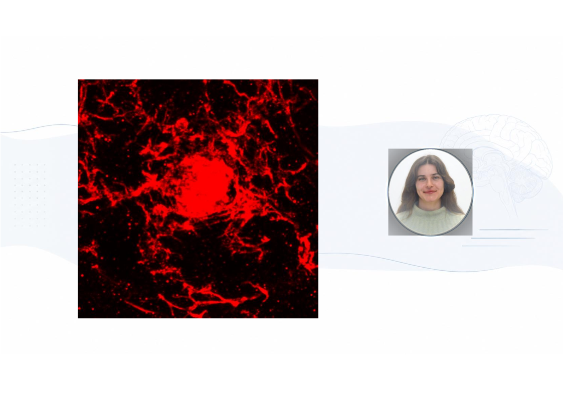
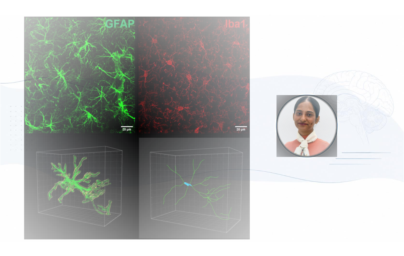
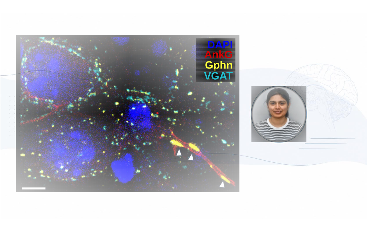
GABAergic neurotransmission primarily mediates inhibitory activity in the central nervous system. These inhibitory neurons are well-known to play a role in physiological processes including brain plasticity, sensory processing, stress reactivity, memory consolidation, and attention. Several post-mortem studies also implicate that GABAergic circuits have been linked to most psychiatric disorders, including schizophrenia, autism, bipolar disorder and major depressive disorder. In order understand the role of GABAergic synapses in affective disorders and to potentially target them therapeutically, it is first necessary to identify the molecular composition of the GABAergic synaptic machinery, particularly the molecules that govern the localization and function of GABAARs. GABAA receptor, a pentameric ligand-gated chloride channel whose synaptic clustering is mediated by the scaffolding protein gephyrin (Gphn). Trans-synaptic adhesion molecule neuroligin-2 (Nlgn2) bridges the pre and postsynaptic compartments by interacting with presynaptic neurexins. Nlgn2 stabilizes the inhibitory postsynaptic density by binding the scaffolding protein gephyrin (Gphn) through its intracellular binding motif, while its proline-rich region interacts with collybistin. Notably, variants in Nlgn2 are associated with a spectrum of psychiatric phenotypes including anxiety and schizophrenia, and Nlgn2 knockout (KO) mice cause prominent anxiety behavior. Additionally, previous research from our group demonstrated that the anxiety phenotype in Nlgn2 KO mice is accompanied by the development of significant Gphn aggregation, raising the intriguing question of whether Gphn aggregation might contribute to the pathophysiology of Nlgn2 variants. The goal of this project is to integrate molecular, cellular, and proteomic approaches to comprehensively investigate the Gphn aggregation. This includes defining the distribution and cellular localization of aggregates within brain regions, identifying interacting partners associated with aggregates under physiological conditions using proximity-labeling strategies such as TurboID, and pinpointing specific site(s) within the protein that are responsible for aggregation through targeted mutagenesis. Together, these approaches aim to elucidate the molecular mechanisms underlying aggregate formation and the interaction networks.

Consequences of aircraft-related noise and air pollution on brain pathophysiology

Yami George, M.Sc.

Aircraft-related pollution is one of the major health-related issues of our time. It mainly consists of ultrafine particles (UFP) and high-intensity intermittent noise. Both factors are linked to the aetiology of cardiovascular and neurological disorders, and in combination, they can lead to stress responses and neurodegenerative disorders (Basner et al., 2014; Münzel et al., 2017, 2023). UFPs with diameters < 100 nm can bypass pulmonary clearance mechanisms, translocate into the bloodstream, and cross the blood–brain barrier, thereby triggering neuroinflammation, oxidative stress, and vascular dysfunction (Block & amp Calderón-Garcidueñas, 2009; Woodward et al., 2015). Additionally, studies indicate that UFPs can directly translocate to the central nervous system via the olfactory pathway, providing a route of entry that bypasses the lungs (Oberdörster et al., 2004). Chronic exposure to aircraft noise elevates stress hormones such as cortisol and catecholamines and activates the sympathetic nervous system (Hahad, Prochaska, et al., 2019; Münzel et al., 2018). Through persistent activation of the hypothalamic–pituitary–adrenal (HPA) axis and inflammatory signalling, these environmental stressors can impair cerebral perfusion, alter heart–brain communication, and contribute to anxiety, depression, and cognitive decline (Ernst, 2017; Hahad, Kröller-Schön, et al., 2019; McEwen et al., 2016). Despite this evidence, key knowledge gaps remain. While associations are established, the precise molecular and synaptic mechanisms underlying brain–heart axis disruption remain poorly understood. In particular, the roles of inflammatory pathways and BDNF/TrkB signalling have not been systematically addressed. Moreover, it is unknown how combined exposure to UFP and noise affects at-risk populations such as individuals with Alzheimer’s disease, diabetes, or hypertension, or whether early developmental exposure produces long-term consequences. This project, conducted jointly by the Institute of Anatomy and the Department of Cardiology at Johannes Gutenberg University Mainz within the MARKOPOLO consortium, will address these gaps. MARKOPOLO is an EU-funded interdisciplinary network that integrates experimental, clinical, and computational approaches to uncover how traffic-related noise and air pollution drive cardiovascular, pulmonary, and brain disorders through shared mechanisms. Using controlled exposure models, we will combine advanced confocal imaging and 3D image analysis with behavioural, electrophysiological, molecular, and activity-mapping approaches to investigate neuroinflammation, synaptic alterations, and neuronal network function in wild-type and disease-model mice. Ultimately, the study aims to define mechanistic links among aircraft-related pollution, synaptic dysfunction, and brain–heart axis pathology, and to identify potential intervention targets for vulnerable populations.